by Joseph Vacca
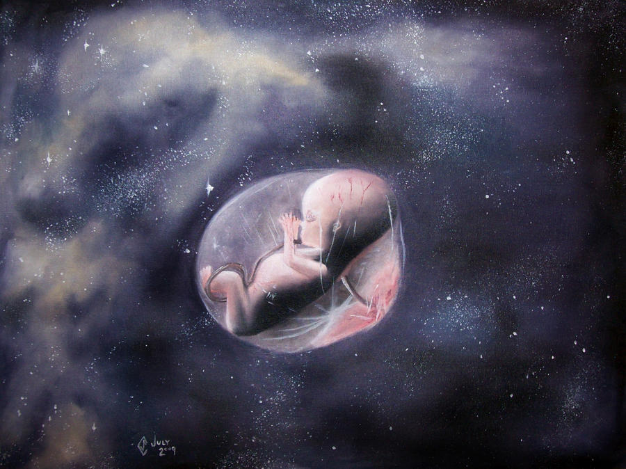
Have you ever really sat down to think about space, that
final frontier? Have you ever wondered if one day you may live to see the human
colonization of another planet; or maybe even recognize the first child born
and raised in space? Well, for all of you active imaginative/sci-fi loving
people out there, I am here to give you the low down on the development of
space children.

Embryo Development
and baby’s first steps…
May not actually be possible… Sorry to start off on a
downer, but it is true. The problem is with fertilization. In cow semen, the
cytoskeletal composition that allows for the tail to correctly generate the
force needed for propulsion is affected by low gravity environments [1]. It may
not have the necessary power to be able to push into the zona pelucida of the
egg. However, studies by NASA scientists have also found that the enzyme that
phosphorylates the tail and causes motion also acts in a hyper activated state
when at low gravity [2]. This means that, although sperm are more motile in
space, they are also functionally unable to fertilize a viable oocyte.
Birth is quite another problem, without gravity to help expel the baby, those present will instead have to pull the baby out of the womb. And could you imagine the amount of free floating liquid, gross. As soon as the baby is out, it misses out on its first lesson, orientation.
In the inner ear there are two gravity-sensing areas, the
saccule for vertical orientation and the utricle for horizontal orientation.
These areas have hair cells within them that are surrounded by little crystals
that are known as otoliths. On earth, gravity pulls these crystals down in the
inner ear, thereby bending the hairs of the sensory cells downward.

Try not to bend over backwards thinking too hard about this one...
The sensory hairs can be bent in any direction, and the
brain interprets the specific bending as specific orientations. However, in
space fluid and objects are free floating; therefore, the otoliths cannot
contact the hairs quite as well, leading to a loss in the perception of tilting
[3]. This will interfere with the baby’s ability to distinguish up from down or
movement from side to side. If ever returned to gravity the child would neither
be coordinated nor balanced. This is because the neural pathways of the child
have not developed to move the body in relation to gravity's pull.

They would probably end up looking something like this on
earth
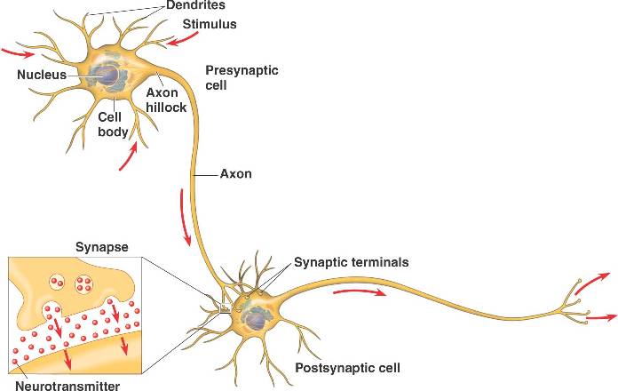
Neural development
Have you ever heard of muscle memory when talking about a
sport or repetitive activity? The same kind of “training” of your muscles must
occur during the first few months of birth [4]. Rats born and raised for 16
days at extremely low levels of gravity never learn how to correct their
orientation when placed on their backs. It was never necessary before. So when
scientists brought the mice back to Earth and placed them on their backs, they
were unable to “properly” right themselves by flipping their legs underneath
them [4]. The weirdest thing about this discovery is that they continued to
improperly turn over for up to a month after returning to Earth! To even
further investigate the effect, mice neurons were stained to observe their
presence and morphology. And just as expected, there was a lower number of
motor neurons branching from the spinal cord to the muscles in the medial part
of the body – these neurons are involved in righting the body’s orientation.
This suggests that the rat’s motor system was biased towards adaptation to a
totally different environment!

Life Alert doesn’t seem so bad now, does it?
These muscles had become used to the weightlessness of
space. But 60% of our muscles are skeletal weight-bearing muscle, meant to hold
our bodies up against the force of gravity [5]. You are using them just to stand/
sit while reading this. So what does all that muscle do if it has no use in
space? Well not only do you not grow the same muscle size and composition as
you would on Earth, but you also have weak branching of the nerves into the
muscle cell as seen in rats [5]. This leads to a decrease in overall
developmental function that cannot be recovered once returned to normal gravity
conditions.
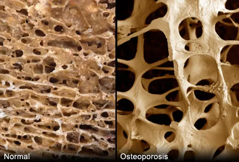
Muscle and Bones
Bones require constant stress in order to develop properly.
Normally, in Earth’s gravitational pull this isn’t too much of a problem.
However, when astronauts are exposed to long periods of time in space, their
bones tend to atrophy, and appear similar to osteoporosis patients [6].

 In developing children this could be even worse. Such soft
bones would easily break and heal incorrectly. These bones would form much like
bone formation in the disease, Rickets [7]. Rickets is formed by a lack of
vitamin D in the diet, thereby causing poor bone formation. A child grown up is
space may look like this:
In developing children this could be even worse. Such soft
bones would easily break and heal incorrectly. These bones would form much like
bone formation in the disease, Rickets [7]. Rickets is formed by a lack of
vitamin D in the diet, thereby causing poor bone formation. A child grown up is
space may look like this:
Citations:
[1] Miller, K. (2002). Floating Fertility. NASA. Available at http://www.nasa.gov/vision/earth/livingthings/floating_fertility_prt.htm
[2] Tash, J.S., Kim, S., Schuber, M., Seiber, D., and
Kinsey, W.H. (2001). Fertilization of sea urchin eggs and sperm motility are
negatively impacted under low hypergavitational forces significant to space flight.
Biol Repro 65(4):1224-31.
[3] Clement, G., Berthoz, A., Bernard, C., Moore, S.,
Curthoys, I., Dai, M., Koizuka, I., Kubo, T., and Raphan, T. (2003). Perception of the Spatial Vertical During
Centrifugation and Static Tilt. In
The Neurolab Spacelab Mission: Neuroscience Research in Space (ed. J.C.
Buckley and J.L. Homick), pp. 5-10. Huston, NASA.
[4] Kalb, R., Hillman, D., DeFelie, J., Garcia-Segura, L.M.,
Walton, K.D., and Llinas, R.R. (2003). Motor System Development Depends on
Experience: A microgravity study of rats.
In The Neurolab Spacelab Mission: Neuroscience Research in Space (ed.
J.C. Buckley and J.L. Homick), pp. 95-103. Huston, NASA.
[5] Riley, D.A. and Wong-Riley, M.T.T. (2003) Neuromuscular
Development Is Altered by Spaceflight.
In The Neurolab Spacelab Mission: Neuroscience Research in Space (ed.
J.C. Buckley and J.L. Homick), pp. 105-109. Huston, NASA.
[6] NASA Science – Science News: Space bones. (2001). NASA.
[March 4, 2014]. Available at http://science1.nasa.gov/science-news/science-at-nasa/2001/ast01oct_1/
[7] Bricker, N.S. (1979). Life beyond the Earth's
Environment: The Biology of Living Organisms in Space. Washington: National
Academy of Sciences. Print.
Pictures:
http://www.theinsidetrainer.com/wp-content/uploads/rickets-vitamin-D-deficiency.jpg


