Monday, December 30, 2013

How to Blog

Greetings, BIO 502ers! 

greeniveyd.com

I hope that your first quarter at Cal Poly was all you hoped for. I am excited to welcome you to winter quarter, when you should be developing your thesis project and establishing a committee (regular MS students) and working your butt off in coursework. Part of that coursework is BIO 502, enigmatically named The Biology of Organisms. This quarter, this means animal physiology, which I know gets you as excited as...


In BIO 502 you are required to write two blog posts. The due dates vary by student and are on the syllabus. The blog posts should be entered on this class blog site, should be about topics that interest you (one rule: they must be about physiology/behavior but can be on any organism), and should be high quality. I will let you figure out what that means on your own, but here are some tips:

1. Check out some of last year's blogs by looking at the archives. Another great example of a blogging graduate student can be found here. Click here for an article on how to write a good science blog.

2. Watch out for the three biggest mistakes that newbie science bloggers make! These are: blog is too short, blog is too long, blog is too technical. These last two are especially sucky, as they can lead to...

illustrationsource.com

2. Really good blogs should have lots of links and images, and not too much text. This will hopefully result in...

myrealtyold.rzware.net

3. Obviously, all blog entries must be 100% original material, written only by you, with citations and image credits where necessary. Please do not use an assignment you have previously completed for your blog post.

4. Read the other students' blogs as they are posted. This will prevent you from posting on the same topic, and besides, info from the blog posts will be on the exams. Please advertise your blog posts and your classmates' on Twitter, Facebook, etc.

DON'T FORGET, THE CLASS HASHTAG IS #Physiologizing

5. Choose your topic carefully! Remember, if you are bored writing your blog post, your readers probably will be, too. You get to pick the topics, so you have no excuse for being boring! Just remember this and you will do fine...


Tuesday, March 12, 2013

Benefits of Reduced Water

Michael Wade


About a week ago, one of my friends posted a video on Facebook that started a big debate and lead to this post. In the video, this guy was advertising Kangen water and it’s filtration system. The guy wasn’t scientifically literate, saying things incorrectly about chemistry and biology that he should have learned in high school. The claim that I got in a debate about was that drinking alkaline water is good for you because it’s literally putting millions and millions of electrons into your body to fight off oxidation. Made me laugh. I told my friend to drink lye (NaOH), find a literature article to support the claims and get back to me. Well… he didn’t drink lye, but he did give me some literature, which prompted my interest in this subject, and will hopefully get some wheels turning in your heads too.

The terms electrochemically and naturally reduced waters are probably foreign to many people. Electrochemically reduced water (ERW) is electrolyzed water produced near the cathode, usually platinum coated, during electrolysis and tends to be alkaline, with a pH of 8-10 (Fig.1). ERW are high in H2 gas and is hydrogen molecule-rich.  Naturally reduced water (NRW) is water that naturally has high levels of H2 gas and is hydrogen molecule-rich. NRW reserves have been found in some parts of Japan and Mexico.


Figure 1. Preparation of electrolyzed water (A), and Chemical reactions at the surface of the platinum cathode (B). (Shirahata et al.2011)

A lot of research is going on in Japan on the benefits of ERWs and NRWs. Much of the research is focusing on diabetes, but there are many other disease models being investigated including uses as an anti-neurodegenerative and anti-cancer drug. This blog will focus mainly on the effects of reduced water and diabetes both in vitro and in vivo.

Type 1-diabetes is a chronic lifelong disease characterized by too much glucose in the blood caused by damaged or nonfunctional b cells. In mouse models, diabetes can be induced with certain drugs whose meachanism is not fully understood, but acts through the production of reactive oxygen species and selectively targest pancreatic b cells. Li et al. induced Type 1-diabetes in a hamster cell line with the diabetogenic drug alloxan. The b pancreatic cells were incubated with different waters prior to exposure to alloxan, then tested for viability. The ERWs and NRWs showed an increase in viability compared to the control after exposure to alloxan(ultra pure water) (Fig.2). Since alloxan increases ROS in b  cells, it is believed that these ERWs and NRWs (from here on will be refered to as RW) exhibit their protective effect via their ability to scavenge and neutralize ROS, which is supported by previous studies showing an antioxidant effect of RWs. Glucose induced insulin secretion was also increased in RW treated groups. Li, et al. hypothesized that this is due to an increase in glucose sensitivity because RW did not increase insulin secretion without glucose stimulation.

As we all know, success with in vitro experiments doesn’t mean success with in vivo experiments. However, with the case of diabetes and RW, in vivo animal models are working and clinical trials are currently being performed. In genetically diabetic mice (db/db), Kim et al. showed that RW significantly lowered the blood glucose levels and raised the insulin levels. In the db/db mice, the size of the pancreas was significantly smaller than that of the control, and interestingly enough, RW treatment increased the size of the pancreas, which is probably why the insulin levels increased.

Type 2-diabetes mice models are also showing success. Jin et al. show that OLETF (type 2-diabetic mice) that blood glucose levels are consistently lower than control mice. One characteristic of type 2-diabetes is hyperlipidemia. OLETF mice treated with RW had significantly lower levels of cholesterol and triglycerides in the blood. Not only do RWs protect against diabetes, but they also protect against diabetic-related complications such as heart disease. GOT and GPT are amino transferases that are secreted into the blood from damaged heart cells. This damage has been linked to lipid deposition the coronary artery causing a blockage, leading to oxygen deficiency. In RW treated mice, the concentration of secreted GOT and GPT were lower than those in the control which is expected due to lower blood lipid levels.


Figure 2. Effect of Tap water vs. RW in Type 2 diabetic mice after an intraperitoneal injection of glucose. (Shirahata et al.2010)

Although the exact mechanism of how RWs treat diabetes, Shirahata et al. have started to fill in some gaps in the insulin cascade. RWs promote phosphorylation of the B-subunit of the insulin receptor via suppression of redox-sensitive tyrosine phosphatases. RWs also activate Akt, a PI3 kinase, which is required for translocation of the glucose transporter, GLUT4, to the plasma membrane. Akt also plays a role in lipid metabolism; however, nobody has looked specifically at that mechanism due to a greater interest in glucose metabolism.

In Japan, there have been a few clinical studies that have shown promise for diabetic patients. An a study with 411 type 2-diabetes, 45% of patients who drank RW showed significantly lower levels of blood glucose, blood cholesterol, LDL (bad cholesterol) and creatine and higher HDL 6 days. A 
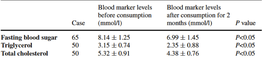
similar study of 50 patients over a two month period showed 89% of patients had a significant decrease in blood glucose, 92% showed a significant decrease in blood triglycerides and total cholesterol levels. Although these two studies weren’t double blind, two double blind studies were conducted later and showed very similar results.


Table 1.  Suppressive effects of RW on 65 diabetic patients and 50 hyperlipidemia patients. (Shirahata et al.2010)


Patients in end-stage renal disease suffer T-cell damage caused by oxidative stress, leading to T-cell apoptosis; patients also have and a low cytokine level and the ratio of Th1/Th2 is not normal. Due to its antioxidant properties, RWs were investigated as a possible treatment in 42 end-stage renal disease patients compared to 12 healthy individuals. After one year of RW treatment, there was a significant increase in the amount of T-cells, a significant decrease in T-cell apoptosis, and the intracellular levels of cytokines was significantly increased, and the ratio of Th1/Th2 was returned to normal levels.


Figure 3. Variety of functions of RW. (Shirahata et al.2011)

On one last note, if diabetes treatment didn’t keep your interest, then maybe this will. RWs have been shown to reduce ethanol-induced hangovers in mice by significantly increasing alcohol dehydrogenase and acetaldehyde dehydrogenase in liver tissues. Although I don’t know of any clinical trials for this, there probably would not be a shortage of volunteers in the college community who wouldn’t want to test this...

RWs are showing great therapeutic potential in a variety of diseases. One of the hallmarks of RWs is that it is completely safe and has zero side effects. Since this “miracle water” has been shown to act as a therapeutic agent for many diseases in Japan, it makes you wonder why there’s almost no research on it here in America. Part of me can’t help but think that the big pharma doesn’t want to make this research known because of the profits they make from disease…


References:

Park, S., Qi, X., Song, S. et al. Electrolyzed-reduced water inhibits acute ethanol-induced hangovers in Sprague-Dawley rats. 2009. Biomedical research. 30(5): 263-269.

Shirahata, S. Hamaski, T., and Kiichiro Teruya. Advanced research ofn the health benefit of reduced water. 2011. Trends in Food Science & Technology. 23: 124-131.

Li, Y., Nishimura, T., Teruya, K., et al. Protective mechanism of reduced water against alloxan-induced pancreatic b cell damage: Scavenging effect against reactive oxygen species. 2002. Cytotechnology. 40: 139-149.

Nakayama, M., Nakano, H., Hamada, H., et al. A novel bioactive haemodialysis system using dissolved dihydrogen (H2) produced by water electrolysis: a clinical trial. 2012. Nephrology Dialysis Transplantation. 25:3026-3033.

Kim, M., Jung, K.H., Uhm., Y.K., et al. Preservative effect of electrolyzed reduced water on pancreatic b cell mass in diabetic db/db mice. 2007. Biological and Pharmaceutical Bulletin. 30(2): 234-236.

Jin, D., Ryu, S.H., Kim, H. W., et al. Anti-diabetic effect of alkaline reduced water on OLETF rats. 2006. Bioscience Biotechnology and Biochemistry. 70(1): 31-37.

Huang, K., Hsu, S., Yang, C., et al. Electrolysed-reduced water dialysate improves T-cell damage in end-stage renal disease patients with chronic haemodialysis. Nephrology Dialysis Transplantation. 25: 2730-2737.

Kicking Kinetosis


Kicking Kinetosis

By: Lesley Stein


It was a dark, chilly, and windy morning in November.  I arrived at Morro Bay to fish for the Collaborative Fisheries Project.  The night before, I checked the swell report, as I am prone to motion sickness, or kinetosis, and unfortunately, it appeared as though rough seas were ahead.  But, with high hopes of somehow overcoming my seasickness with a motion sickness pill and some ginger root tablets, I boarded the boat the following morning.  After departing from the peaceful bay, I began to feel a little warm inside. My mouth began to salivate and my body began to sweat while my stomach started churning, and I was extremely nauseous.  My motion sickness had come back with a vengeance.  All I wanted to do was lay down and close my eyes, until the boat docked.  Not only was this desire to remain unsatisfied, but I furthermore still had to tag, weigh, measure and release the catch soon to be reeled in.  My miraculous concoction of over-the-counter motion sickness medication and ginger tablets did not live up to my expectations and without a moment to find a trash can or even a fishing bucket, I rushed to the side of the boat and, well, fed the fish.   Although I knew everyone was disgusted watching me hurl over the side of the boat every five minutes, I must say, they were highly impressed with my multi-tasking skills of projectile vomiting while gripping a flailing 20-pound lingcod in my hands! 


How and why does this happen?
So what role does the inner ear (vestibular system) play in all of this, you ask?  Well, just as, your muscles, eyes, and skin receptors detect motion or a lack of motion, so too, does your inner ear.  The vestibular system (see Figure 2) consists of a semicircular canal system, conveying rotational movements, and otoliths, expressing linear accelerations.  Within the semicircular ducts are tiny hair cells called crista, which detect the movement of fluid inside the ear.  


In a nutshell, it is caused by the inner ear (see Figure 1). Overstimulations of the labyrinthine canals of the ear initiate motion sickness.  Motion sickness occurs when the inner ear, the eyes and other parts of the body that distinguishes motion, send contradictory messages to the brain.


http://www.directhearingaids.co.uk/index.php/33/how-hearing-balance-work-together/ 

Figure 1. The three main parts of the ear: external, middle, and inner.  The inner ear plays a role in motion sickness.

When we move our bodies intentionally, for example, when we run or jump, the input from all three of these pathways to our brain is in sync.  However, when we experience unintentional movement, as occurs when aboard a moving fishing vessel, the input to the brain does not coordinate.  During motion sickness, the brain receives conflicting messages from these numerous interpreters that express contradictory states of the body’s physical status.  For example, on a boat, your feet are not physically moving, so your joint sensory receptors say to your brain, “I am still.”  However, your eyes perceive the vanishing shoreline and flowing wake behind the boat indicating movement, telling your brain, “I am moving.”  Similar to reading a book in the car—your eyes are focused on a stationary page, while your body registers the movement of the car.  And your skin receptors feel wind, as experienced during a fast paced run, telling your brain, “I am moving.”  But your legs and arm muscles are not moving, thus tell your brain, “I am still.”  In other words, you feel motion, but you don’t see it.  Or, you see motion, but you don’t feel it.  Imagine how you would feel trying to decode these conflicting messages!


https://rdl.train.army.mil/catalog/view/100.ATSC/C696BACA-168F-4B4A-9750-BB445E08BECE-1300757497629/3-04.93/chap9.htm 
Figure 2. The anatomy of the inner ear.


Figure 3.  (a) The head is positioned straight, the, therefore the cristae are also straight.  (b)  When the head turns the cristae flex in the opposite direction.  Note: fluid inside the inner ear is moving the cristae. 


Motion sickness occurs when our brain receives these contradictory messages.  My personal encounter with motion sickness was specific to seasickness, which is just one type of motion sickness.  Many people experience the symptoms of motion sickness traveling in a car, train, airplane, bus, subway or while watching video games, IMAX movies, or riding roller coasters.  While I was on the boat, my inner ears and skin receptors sent signals to my brain indicating motion.  However, my muscle receptors registered my motionless status as I was standing still, or at times, sitting still.  My eyes were glued to the floor that wasn’t moving, but as soon as my eyes met the horizon or shoreline, my eyes sent an opposing signal indicating motion—conflicting with the one sent just moments ago from the same receptor. 

This mismatch of neural signals ultimately, activates the postrema in the brain, which controls emesis (vomiting), as illustrated in the figure below (Figure 4).  The vomiting-reflex is controlled in the vomiting center, or the lateral reticular formation, located in the medulla oblongata at the base of your brain.  The link between motion sickness and vomiting is not wholly understood, but one theory includes the toxin theory.  According to this theory, vomiting is a defense mechanism against neurotoxins.  Due to the conflicting signals sent to the brain, the brain interprets one of them as a hallucination, triggered by poison or drugs.  The brain, in turn, instructs the body to purge itself of these toxins through emesis.


(https://www.healthtap.com/#user_questions/220991-why-do-we-throw-up-when-we-get-motion-sickness)
Figure 4. The pathway of the postrema area in the brain, controlling vomiting

So, we now know why we get motion sickness, but what do we do about it?  

There are several different remedies to help alleviate the symptoms, but there is no “cure” per sue.  One of the most popular methods, includes the one that failed to come to my rescue that Saturday morning, over-the-counter medication.  These include drugs like Dramamine or Bonine.  These are antiemetics, or drugs that inhibit vomiting.  These drugs work by blocking the messages of certain neurotransmitters like serotonin, acetylcholine, dopamine, and histamine.  If the brain does not receive information from these neurotransmitters, it is unable to respond.  These antiemetics block the transmission of information from the vestibular apparatus to the lateral reticular formation (vomiting or emetic center) in the medulla oblongata.   See the figure below (Figure 5) to see the where the antiemetics can block neurotransmitters’ signal to the vomiting center.  


Figure 5. Different pathways of antiemetics blocking neurotransmitters' signals in the vomiting center


But these antiemetics also tend to pack another punch—extreme drowsiness.  The reason being is because some antiemetics are also antihistamines, like Dramamine.  The main ingredient in antihistamines that causes drowsiness is called diphenhydramine that fights histamine (as seen above) by binding to the receptors of specific cells in the body that histamine would typically bind to otherwise, blocking them from relaying their message.   If histamine does not have access to these receptors, it cannot react on the body.  But antihistamines are not solely responsive to histamine receptors, but acetylcholine receptors as well.  If diphenhydramine clocks an acetylcholine receptor, acetylcholine reuptake no longer occurs, causing an excess of acetylcholine, and thus drowsiness.  The only medication that I have taken (and I have take a lot) that did not cause this unfavorable side effect is Triptone, which is unfortunately, no longer being manufactured. Scopolamine patches, placed behind the ear, as well as, wristbands applying pressure to specific pressure points believed to alleviate motion sickness, has been successful for many.  Ginger root pills or chews can also diminish the side effects of motion sickness, phenols in ginger also help relax the stomach muscles, including the pyloric valve; therefore, reducing the activity of the stomach.  Ginger also promotes the stomach to secrete digestive juices or enzymes, furthermore, neutralizing stomach acids. Other than these more medicinal aids, a few words of personal advice...Always have food in your stomach before you travel, it may help you not get sick and if you do get sick, at least you will not be throwing up bile..  And lastly, carry a barf bag to avoid some embarrassment.


References:


Bles, W., J.E. Bos, B.D. Graaf, E. Groen, and A.H Wertheim. 1998. Motion sickness: Only one provocative conflict? Brain Research Bulletin 47: 481-487.

Brad Bowins. Motion sickness: A negative reinforcement model. 2010. Brain Research Bulletin 81:7-11.

Charles Oman. 1998. Sensory conflict in motion sickness: an observer theory approach. NASA, Ames Research Center, Spatial Displays and Spatial Instruments 16-54.


Huang, Y.D., S.W. Xia, P. Dai, and D.Y. Han. 2011. Role of AQP1 in inner ear in motion sickness. Physiology & Behavior 104: 749-753

Karen Naifeh. 1986. Exploratory Studies of Physiological Components of Motion Sickness: Cardiopulmonary Differences Between High and Low Susceptibles. National Aeronautics and Space Administration ARC 275a.


Theeuwes, J., A.F. Kramer, S. Hahn, and D.E. Irwin. 1998. Our eyes do not always go where we want them to go: capture of the eyes by new objects. Psychological Science September 9: 379-385.


Images:


(http://thetravelingtype.files.wordpress.com/2011/08/seasick.jpg)


(http://www.directhearingaids.co.uk/index.php/33/how-hearing-balance-work-together/)


(https://rdl.train.army.mil/catalog/view/100.ATSC/C696BACA-168F-4B4A-9750-BB445E08BECE-1300757497629/3-04.93/chap9.htm)


(http://humanphysiology2011.wikispaces.com/10.+Sense+Organs)


(https://www.healthtap.com/#user_questions/220991-why-do-we-throw-up-when-we-get-motion-sickness)


(http://www.nes.scot.nhs.uk/prescribing/topics/palliativecare/page13.htm)