Thursday, January 29, 2015

Physiology of Sex

By Aubrey Stiers

Physiology of Sex


No not that kind of sex! Differences in brain physiology between sexes, i.e. men and women. Now before you get too disappointed and leave this blog you should know that there are important sex differences in brain structure, function, and chemistry that developed early in your life and that have an impact on everything from your relations to your metabolism of drug. There are many variables that contribute to brain development and structure and I'm not going to argue one side or the other of the nature vs. nurture debate, but rather provide you with interesting and informative scientific data about the physiological differences in the brains of men and women.  
Let's start with a story. Imagine a woman struggling to sleep. Let's say she has two wild toddlers driving her crazy all day or mountains of homework loading her down. Her doctor finally prescribes Ambien to help her "fall asleep fast, stay asleep longer, and wake rested and refreshed"(8). However, after taking Ambien she experiences hallucinations and amnesia, sleepwalking, sleep driving and sleep eating, and even has sex while totally asleep (7). Her husband, who does not mind this last side effect, also takes Ambien but does not experience any of these side effects. What could cause such a drastic difference in side effects between this husband and wife?


In January of 2013, the FDA released a statement in which it recommended that women reduce the dose of Ambien and other sleep aides by half (3,2). The reason behind this sudden announcement? It was discovered that women metabolize the main active ingredient in Ambien much slower than men, half as fast in fact. This means that women were waking up with large amounts of Ambien still in their systems putting them at risk for accidents due to impaired function (2). The sleep studies conducted using Ambien did not include females due to the thought that fluctuations in (menstrual) hormones would make it "more difficult to draw conclusions from their findings, and any findings in male study subjects translated to females" (2). This is just one example in which sex differences in physiology significantly influenced the effects of a drug. “Sex differences exist in almost every chronic disease. For example, cardiovascular disease, Alzheimer’s, autoimmune disorders such as rheumatoid arthritis and multiple sclerosis, depression, anxiety disorders, autism, schizophrenia…the list goes on"(4), says Dr. Jill Goldstein of Harvard Medical School. It can no longer be denied that sex differences in the brain are important.


This recent FDA announcement brings up many of questions. For instance, how does the male and female brain differ? When does this differentiation occur in development? Is there really a physiological reason for my girlfriend's craziness? There may be answers for all of these questions, except for the last one.  
           
 Differentiation of the Brain


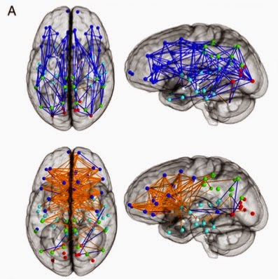
Top showing greater intra-hemisphere connections in the
male brain and the bottom showing more
inter-hemisphere connections in the female brain.


There are many similarities between the differentiation of the brain and reproductive systems. For example, both differentiation processes begin in utero. "[S]ex divergence typically occurs in brain development around the eighth week of gestation"(4) and it is known that "steroids act early in development to dictate [both] adult sexual behavior and brain morphology"(6). In a sample of 949 youths aged 8–22 years, imaging of brain connectivity revealed that there are "unique sex differences in brain connectivity during the course of development" (5). The results show that "males had greater within-hemispheric connectivity", as well as enhanced organization, compared to females who had higher inter-hemispheric connectivity and cross-lobe integration (5). 

Analysis on the child (B), adolescent (C), and young adult (D) groups is shown. Intra-hemispheric connections are shown in blue, and inter-hemispheric connections are shown in orange.
So it seems that the description of men having brains full of organized boxes (my sex-ed teacher referred to this as the "waffle brain") is an accurate description of the organized physiology that is "optimized for communicating within the  hemispheres" (5). Alternatively, the female brain, AKA the "spaghetti brain", is less organized but establishes connections with more parts of the brain. This trend is also seen in developing children with boys "displaying higher intra-hemispheric connectivity [than girls] of the same age" (5). 

These physiological structures suggests that the male brain is "structured to facilitate connectivity between perception and coordinated action," whereas female brains are organized to "facilitate communication between analytical and intuitive processing modes"(5). While males typically have a larger body mass than women, they also have "larger crania ... and a higher percentage of white matter" that contains myelinated neural axons and cerebrospinal fluid (1). Women on the other hand have a higher percentage of gray matter that is made up of neural bodies(1). A review found that "In men, IQ correlates with gray matter volume in the frontal and parietal lobes; whereas in women, IQ correlates with gray matter volume in the frontal lobe and Broca’s area,[a region of the frontal lobe associated with speech production] ... suggesting that men and women use different brain areas to achieve a similar IQ"(1). 

Interestingly, this physiological study was also paired with a behavioral study on the same 949 children and found pronounced sex differences. "[F]emales outperform males on attention, word and face memory, and social cognition tests and males perform better on spatial processing and motor and sensorimotor speed"(5). So it appears there is a physiological reason for the differences between men and women. 
Did someone say shoe SALE?!!
Let's get it on.... oh wait the games on! 

So how do differences in brain physiology explain the difference in metabolism of Ambien? In a study measuring cerebral blood flow, the results showed that women have higher global cerebral blood flow than men both while at rest and during cognitive activity however the overall cerebral metabolic rate of glucose was equivalent between the two sexes (1).While metabolism of drugs was not evaluated, this data suggests that any drug that can cross the blood-brain barrier is better distributed in the female brain, which is an important feature when studying drug dosage. Greater distribution of the drug means that it may take longer to metabolize the drug. Studies that examined the sex difference in neurotransmitters found that women have higher concentrations of serotonin in the blood relative to men. Why does that matter you ask? Because serotonin is a possible cause of or contributor to mood disorders, sleep and eating disorders, and schizophrenia(1). "Sex differences in [serotonin] function may underlie the known gender difference (women > men) in the prevalence of depression and may impact pharmacological treatments that target [serotonin] neurotransmission"(1). These structural, functional and chemical differences between the male and female brain illustrate the need to better understand their influence on neuropsychiatric disorders, drug dosing and metabolism and may even help you better understand your partner because of a better understanding of his/her brain. 

Still have questions about your partner's brain and behavior? Or do you just want more entertainment? I suggest you watch the video below that offers a comical explanation of the difference between men and women's brains. 


References:
Images and videos in order of appearance:
1. http://www.medindia.net/news/three-scientists-awarded-the-nobel-prize-for-physiology-142198-1.htm
2. http://middlesexhospital.org/our-services/hospital-services/the-comprehensive-sleep-center/services/other-sleeping-disorders
3. http://perrishillspharmacy.com/tags/public-service-announcement 
4. http://philbasiceducation.blogspot.com/2013/02/girls-and-science.html
5. http://philbasiceducation.blogspot.com/2013/02/girls-and-science.html
6. Gungor, Mark. "A Tale of Two Brains." Online video clip. YouTube. YouTube, 28 Feb 2011. Web. 24 January 2014. <https://www.youtube.com/watch?v=3XjUFYxSxDk>.

In text citations in alphabetical order: 
1. Cosgrove, K. P., C.M. Mazure, J.K. Staley. Evolving knowledge of sex differences in brain structure, function and chemistry. 2007. Biological Psychiatry 62:847-855.
2. Curley, Allison. “His and Hers: Sex Differences in the Brain.” Brainfacts.org. Society for Neuroscience. 7 May 2014. Web. 24 Jan. 2015. <http://www.brainfacts.org/Brain-Basics/Neuroanatomy/Articles/2014/His-and-Hers-Sex-Differences-in-the-Brain>.
3. “FDA Drug Safety Communication: FDA approves new label changes and dosing for zolpidem products and a recommendation to avoid driving the day after using Ambien CR.” FDA. U.S. Department of Health and Human Services. 14 May 2013. Web. 24 January 2015. <http://www.fda.gov/drugs/drugsafety/ucm352085.htm>.
4. Hart, Erin. “ Sex and the Human Brain.” Brain World. Brain World Magazine. 14 June 2010. Web. 24 January 2015. <http://brainworldmagazine.com/sex-and-the-human-brain/#sthash.rph5c1pi.dpuf>.
5. Ingalhalikar, M., A. Smith, D. Parker, T.D., Satterthwaite, M.A. Elliott, K. Ruparel, H. Hakonarson, R.E. Gur, R.C. Gur, and R. Verma. Sex differences in the structural connectome of the human brain. 2014. Proceedings of the National Academy of Sciences of the United States of America 111:823-828.
6. McCarthy, Margaret M. Estradiol and the developing brain. 2010. American Physiological Society 88: 91-134. Web. 24 Jan. 2015.  <http://physrev.physiology.org/content/88/1/91>.
7. Sommerfeld, J. "While I was sleeping: shopping sprees, sugar binges and other confessions of an ambien zombie." Today.com. NBCnews.com, 13 November 2014. Web. 24 January 2015. <http://www.today.com/health/while-i-was-sleeping-shopping-sprees-sugar-binges-other-confessions-1D80287242>.
8. The Ad Collector 2. "Ambien TV Ad (2003)." Online video clip. YouTube. YouTube, 3 Nov. 2013. Web. 24 January 2014. <https://www.youtube.com/watch? v=LB84dKlbkoE>.

Food to die for


By Evelyn Puspitasari

We’ve had this discussion before, food is good. I personally have always loved seafood. Cooked seafood? Yes. Raw fish, you ask? Yes. I’m always down for sashimi. Any day. Hit me up. Have you heard of fugu though? If you say no, shame on you. Fugu is delish. So, let’s talk about fugu. Fugu is what the Japanese call pufferfish. 

Yep, they're cute.
They are also known as blow fish. That is because when they feel endangered, they can.. blow up.. to a ball of spiky fish. 


Yes, that is a part of their defense mechanism. But that is not their only defense strategy, they also have tetrodotoxin (TTX) coursing through their body, with the highest concentrations in their ovary and liver. Contrary to popular believe, the toxin cannot affect you if you get poked by one of their spikes. It will affect you, however, if you eat the fish and it wasn’t properly prepared. Hence, only certified chefs are allowed to prepare pufferfish. In Japan, where the fish is considered as a delicacy, chef candidates have to go through 2-3 years of apprenticeship, followed by a final examination where they have to take a written test, a fish identification test, and a practical test of preparing and eating the fish. The Japanese take this really seriously, it’s no joke. 

Fugu has to be prepped while the fish is still alive. Not to worry though, chefs severe the spinal cord of the fish to keep the pain level to a minimum. Here's a video of proper methodology to prepare fugu. Do not do this at home. Unless you have a license, which you probably don't.

Despite all the precautions, accidents do happen. Last October in Brazil, eleven people had to be rushed to the hospital when they accidentally ate a puffer fish that was mixed up with the rest of their lunch. Most recently, a brother and sister showed up to a hospital complaining of symptoms that are related to TTX poisoning. Turns out, they were poisoned from the dried fish they bought in New York. Oh, did I mention TTX poisoning has no antidote? Luckily, the victims from both cases all recovered.. Despite having no antidote, victims have a chance to fully recover as long as they seek help in a timely manner. In this case, all they can do is to wait until the toxin has run its course, while being hooked up to a respirator that breathes for them. This is because TTX is a neurotoxin that will paralyze the muscles, causing the victim to have difficulty breathing, and without help will die from asphyxiation. Also, TTX victim remains conscious even when they can’t move or breathe. Like a motionless zombie. 

Why does TTX have these effects on its victims? How does it work? 

Well.. We know that neurons are responsible for communication in the form of electrical signals from one point to another. An action potential is required for a neuron to communicate with other neurons, muscles, or glands. There are three phases of action potential: depolarization, repolarization, and hyperpolarization.
Three phases of action potential
Generally speaking, the membrane potential of the organism must shift to be more depolarized than before, which means the membrane potential will become less negative. Followed by a rapid repolarization of membrane potential to a slightly more negative state compared to its original resting potential (hyperpolarization). The drop in membrane potential is what we call repolarization, that is triggered by the closing of specific ion channels and opening of different ion channels in the membrane. Hodgkin and Huxley, the dynamic duo, did a lot of study with the giant squid’s axon to find out that an action potential begins with an inflow of Na+ through voltage-gated channels that will depolarize the membrane, then an outflow of K+ through voltage-gated channels will hyperpolarize the membrane. 

Understanding the mechanism of action potential was a crucial step in figuring out the TTX mechanism. The next step was to determine the structure of the sodium ion channel which was done by a team in Japan led by Shosaku Numa who used Electrophorus electricus, the electric eels, as their model animal. Their accomplishment tells us that the sodium channel is made up of a single peptide chain with four homologous domains, each consisting of six transmembrane helices.
Narahashi, et. al. made a hypothesis that TTX selectively inhibits the sodium transport mechanism that results in the conduction of nerve and muscle. To prove their hypothesis, they treated giant axons of lobster with TTX at different concentrations of 1x10⁻⁷ to 5x10⁻⁹ g/mL. Voltage-clamping was performed and the result of their experiment shows that there is no increase in influx of Na+ that normally occurs during depolarization when the axon was exposed to TTX, whereas the outflux of K+ was not affected.
Turns out the binding site for TTX consists of amino acid residues that form a pore where Na+ ions come in. When TTX is present, it blocks the pore, thus preventing the entry of Na+ ions through the voltage-gated channel. Hence, resulting in the absence of action potential which means there will be no communication from neurons to nerve tissues or muscles! That is why the victims of TTX poisoning get zombified… 

Pufferfish themselves are obviously not affected by their own toxin. Simple substitutions of amino acid residues in their voltage-gated channels makes them to be highly resistant to their own toxin. Pretty cool defense mechanism, pufferfish. 


References:
Nakamura, Y., S. Nakajima, and H. Grundgest. 1965. The action of tetrodotoxin on electrogenic components of squid giant axons. The Journal of General Physiology 48: 985-996.
Häusser, M. 2000. The Hodgkin-Huxley theory of the action potential. Nature Neuroscience 3: 1165-1165.
Narahashi,T., J.W. Moore, and W.R. Scott. 1964. Tetrodotoxin blockage of sodium conductance increase in lobster giant axons. The Journal of General Physiology 47: 965-974.
Soong, T.W. and B. Venkatesth. 2006. Adaptive evolution of tetrodotoxin resistance in animals. TRENDS in Genetics 22:621-626.
Venkatesh, B., S.Q. Lu, N. Dandona, S.L. See, S. Brenner, and T.W. Soong. 2005. Genetic basis of tetrodotoxin resistance in pufferfishes. Current Biology 15: 2069-2072.
http://en.wikipedia.org/wiki/Fugu

Images:
https://s-media-cache-ak0.pinimg.com/originals/6e/d0/3e/6ed03e0f3f19e36ca7898fc265c1645a.jpg
http://media-cache-ak0.pinimg.com/736x/8d/04/ac/8d04acae13e70c35c1fda76cee1c2591.jpg
http://www.life.umd.edu/grad/mlfsc/zctsim/ionchannel.html
http://www.bio.miami.edu/tom/courses/protected/ECK/CH05/figure-05-26a.jpg
http://chalkandwater.tumblr.com/post/106655012503/a-small-japanese-puffer-fish-is-the-creator-of-one
Freeman, Scott. Biological Science. San Francisco: Benjamin Cummings, 2011. Print.