Thursday, January 31, 2013

The Cephalopod Nervous System: Convergent evolution...or something way cooler than we vertebrates got?!
by Heather Price

Octopuses have long been renowned as the planet’s most intelligent animals lacking a backbone. Their capacity for learning and problem-solving, (and penchant for escaping from tanks and consuming their neighboring aquarium mates), combined with their uniquely flexible, color-changing, eight-appendaged body, make these cephalopods some of the most mysterious and fascinating creatures in the sea.  

The octopus belongs to a group of molluscs called Cephalopods (meaning “head-foot”), which also includes squid and cuttlefish.  There are currently 289 recorded species of octopus, ranging in size from 2 cm (our very own California Lilliput Octopus) to the Giant Pacific Octopus which can reach a diameter of 9 meters!  In nature, the octopus is the master of inventive disguises, both physical and behavioral, that allow it to surprise its prey.  In the lab, octopuses have shown their ability to associate tactile and visual cues with rewards, to navigate mazes, and to teach each other more rapidly than a human researcher can teach an octopus. This high level of intelligence has often caused the octopus brain to be compared to that of vertebrates.

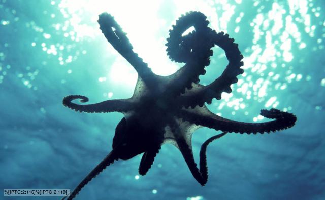
In addition to their intelligence, octopuses also exhibit strikingly rigid, jointed-appendage-like movements, despite the practically unlimited range of flexibility of their arms.  This has also led many people to draw comparisons with the vertebrate nervous system, and to declare that the similarities between the two are an exemplary model of convergent evolution.
While octopuses seem to share more in common with vertebrates than their more closely related invertebrate cousins, the central nervous system of our tentacled friends is actually arranged quite differently than those of backbone-endowed organisms.
The Cephalopod nervous system is divided into three main parts:
1. a central brain surrounded by a capsule made of cartilage
2. two large optic lobes connected to camera-like eyes
3. a peripheral nervous system connected to the arms
The octopus brain is comprised of about 40 lobes, each with its cell body on the outside, and its dendrites arranged into a central mass.  Unlike in most vertebrate brains, stimulation of a particular neuron in the octopus brain does not activate movement of one particular part of the octopus body, but instead triggers an entire series of movements comprising a complex behavior. Researchers have found that octopus arms actually cannot be stimulated individually, and that at least two arms move at a time in response to any one neuronal trigger.
Octopuses has over 500 million neurons, (which is similar to the number our canine companions possess), but unlike Fido, the octopus has about two-thirds of its nerve cells contained within its peripheral nervous system.  Concentrations of ganglia at the base of each arm control most of neuronal processing involved in movement.  The central brain will send an initial message to the peripheral ganglia, which will then coordinate a suite of complex movements. So unlike their human counterparts, male octopuses can legitimately claim that their hectocotylus (a modified arm used for inseminating females) has a mind of its own!
In addition to having arms with eight minds of their own, the octopus nervous system is unique in that it controls color-changing cells in their skin, known as chromatophores.  These chromatophores allow octopuses to rapidly change their appearance in order to camouflage with their environment, or communicate with one another.  Chromatophores consist of a central pigment-containing molecule surrounded by radial muscles that are controlled by the brain. Recent studies have shown that it is actually the rate of expansion and contraction of these radial muscles that determines the pattern on the octopuses’ skin, and not the intensity or type of pigment. While there are several species of vertebrates that exhibit color-changing abilities, none compares in rapidity or complexity to that of Cephalopod chromatophores.
Our tendency as Homo sapiens is to compare anything we regard as highly intelligent to ourselves.  Thus, the frequent comparisons between the octopus nervous system and our own.  However, when one looks more closely at the actual structure and function of the octopus brain and body, it becomes clear the convergence between these clever creatures and ourselves isn’t all that close.  In fact, the octopus may have some adaptations that are much cooler and more complex than we vertebrates could dream of evolving!
Also, in case you’ve been kept up at night wondering whether to say “octopuses” or “octopi” when telling your nerdy science friends about your recent aquarium visit...a Webster’s editor clears it up here nicely:

References:
Gray, E.G., and J.Z. Young. 1964. Electron microscopy of synaptic structure of octopus brain. The Journal of Cell Biology. 1:87-103.

Monday, January 28, 2013

What do Dead Bodies, Nematodes, and a former Oregon State Beavers Pitcher Have in Common?


by Ellen Mintz

          Believe it or not, these three seemingly unrelated organisms have each undergone a process that is incredibly significant in medical history and current scientific research.  To really answer that question though, we need to take a closer look at the most significant and important organ in our body: the brain.
           The brain, along with the spinal cord, comprises the central nervous system. As its name implies, this is the command center of all of the activity that keeps our bodies alive, aware, and functioning.  The brain controls crucial processes like breathing, digesting, and waking up in the morning. Stimuli from the environment around us send messages through our bodies in the form of electrical signals.  Neurons are the cells that transmit these signals, communicating from cell to cell through the release of tiny chemicals called neurotransmitters.  Upon reaching the central nervous system, these signals are processed and delivered as electrical impulses to effectors such as skeletal muscle and digestive organs.
  
            In addition to movement, the brain controls emotions and feelings.  These arise from increased activity in different parts of the brain (check out the map below!).  Regions including the amygdala, frontal cortex, hypothalamus, and cerebral cortex can all lead to feelings of sadness and depression.

Brain Anatomy
            Many tissues around the body such as the skin, are quick to repair themselves following damage from injury or the natural process of aging through the use of unspecialized stem cells that then differentiate into the needed cell type.  The central nervous system, however, is notoriously difficult to repair and was believed to have no source of stem cells. In 1998, scientists discovered cells in the hippocampus that were able to become new neurons, and subsequent discoveries of more of these cells led to the hope that conditions such as paralysis that are often the result of traumatic brain injury could be treated.  This has of yet not been proven to be completely successful.

Fluorescence image of a neural stem cell
            It has taken centuries to accumulate all of the knowledge that we have today about the brain.  Starting in the late 1700s and into 1800s, research in neurobiology and of the electrical impulses in the nervous system made huge strides.  It was during this time that scientists discovered that muscles and nerves were excited by electrical signals.  This revelation was demonstrated by applying electric currents to the bodies of cadavers (dead bodies, anyone?!), resulting in macabre post-death movements sure to excite any pre-movie era horror enthusiasts. Eventually, this led to the discovery by Italian scientist Luigi Rolando in 1809 that the brain responded to electrical pulses also.

            It took another 50 years for scientists to develop the knowledge and instruments to be able to apply this knowledge of the electrical conduction of the central nervous system directly to the organs themselves, and use electricity to control different levels stimulation as a method for investigating brain function.  First in animals like dogs and monkeys, and much later in humans, electrical stimulation of the brain provided knowledge the physiology of this organ.  When this was then applied to the treatment of different disease conditions, particularly mental illness, the fun really began!

What ECT actually looks like!
            One of the first methods of electrical brain stimulation to treat depression and personality and mood disorders was electroshock therapy, now called electroconvulsive therapy, or ECT. The idea behind electroconvulsive therapy is to cause a huge storm of electrical activity in different parts of the brain.  This invokes a small seizure in the patient and resets electrical pathways and brain chemistry.  While often seen fictionalized in popular media as a painful, bone jarring and maniacal spasms, it is in reality done under anesthesia and can be successful when other treatments have failed.
NOT an accurate representation of ECT!

            In 1985, a procedure called transcranial magnetic stimulation, or TMS, was introduced as a less invasive and intense method of mood adjustment and depression treatment and approved for use on humans in 2009. This technique involves placing electromagnetic coils near the skull to deliver quick magnetic pulses to the brain, increasing the firing of neurons in mood associated brain regions.  There is a decrease in the risk of inducing a seizure using this procedure, making it safer and applicable to a wider range of patients than traditional ECT.
            
C. elegans
        Taking the idea of magnetic stimulation multiple steps further, a physicist at SUNY Buffalo, Dr. Arnd Pralle, utilized magnetic fields to heat up nanoparticles placed in neurons in the brain.  These particles activate temperature sensitive ion channels in the cells, causing them to react and potentially stimulate positive changes in neuron firing patterns. Although this method has yet to be tested in human brains, it has been shown to cause Caenorhabditis elegans, a nematode used as a model organism in developmental biology and neurology (a test subject like the dead bodies of earlier…), to change what direction they were moving in. Dr. Pralle and his collaborators are currently investigating using genes from bacteria that are known to make magnetic nanoparticles and integrating them into humans using a viral therapy, in addition to other biocompatible delivery methods.  Ultimately, this technique would be able to elucidate neuronal circuits in the brain by allowing stimulation of different regions controlling mood and behavior, and could be used as a research tool to study conditions such Parkinson’s disease and traumatic brain injury that resulted from dysfunctional or impaired neurons.

            The spinal cord has also been subjected to electrical stimulation as well, however not for the same purposes as the brain.  Electrical stimulation of the spinal cord has been investigated as a way to improve limb function and health, focusing on patients with spinal cords or body parts that have been damaged by accidents or debilitating diseases.  Because the spinal cord transmits electrical signals and impulses from the brain and reflex neurons to muscles and organs around the body, damage can result in loss of movement and control, lack of sensation to stimuli including pain, temperature and touch, and reduced to nonexistent function in crucial organs and systems including the heart, lungs, digestive, and reproductive systems.  It has been shown however that rats with implanted electrodes in their spinal cord regain minor leg function when placed on a treadmill, and eventually are able to walk on their own.  Brief periods of epidural spinal cord electrical stimulation was shown to relieve pain, result in skin ulcer healing, and increase circulation in patients suffering from limb-threatening ischemia.

            Still curious about how baseball fits into this topic? In 2006, Rob Summers, a pitcher for Oregon State, was paralyzed from the neck down in a tragic hit and run accident, ending his baseball career.  Intense rehabilitation and therapy improved his upper body movement and he eventually regained the use of his arms.  In 2009, he sought out the help of Dr. V. Reggie Edgerton, a professor and neurobiologist at UCLA who had previously led spinal cord studies on electrical stimulation in rats.  Dr. Edgerton and his collaborators implanted electrodes in the lower sections of Summers’ spinal cord and through electrical stimulation, were able to temporarily reactive the neural networks in his spinal cord.  Summers was able to move his legs while on a treadmill with upper body support, briefly stand on his own, and also experienced huge improvements in blood pressure, bladder and sexual function, and temperature regulation. 

            All of these therapies involving electromagnetic activity and the central nervous system to treat depression and other diseases and pathologies have many benefits and are good starting points for the development of safer and less invasive procedures.  Further applications of these concepts could provide longer, healthier, and happier outlooks to those suffering from paralysis, depression, and neural diseases like Parkinson’s.  In structures as multifaceted and complex as the brain and spinal cord, it is anyone’s best guess as to from where the next discovery will come!
           
References
Berg, Nate. "Spine Stimulator Lets the Paralyzed Stand Again." Discover. Jan 2013: 36. Print.

Clemmons, Anna Katherine. "Rob Summers Willing to Walk Again." ESPN.com. 24 Dec 2011: Web. 28 Jan. 2013. <http://espn.go.com/espn/otl/story/_/id/7373090/rob-summers-former-oregon-state-beavers-pitcher-paralyzed-accident-never-imagined-impact-world-baseball>.

"Electroconvulsive Therapy." Mayo Clinic. Web. 29 Jan 2013. <http://www.mayoclinic.com/health/electroconvulsive-therapy/MY00129>.
Freedman, David. "The Happiness App." Discover. Jan 2013: 10-11. Print.

Harkema, Susan, Yury Gerasimenko, V.R. Edgerton, et al. 2011. Effect of epidural stimulation of the lumbosacral spinal cord on voluntary movement, standing, and assisted stepping after motor complete paraplegia: a case study. Lancet. 377. 1938-47.

Hsu, Charlotte. "Effort to Remotely Control Brain Cells Gets Push from Innovative Grant." News Center. State University of New York at Buffalo, 16 Apr 2012. Web. 28 Jan 2013. <http://www.buffalo.edu/news/releases/2012/04/13359.html>.
Jacobs, Michael J.H.M., Paul Jornig, Roeland Beckers, et al. 1990. Foot salvage and Improvement of microvascular blood flow as a result of epidural spinal cord electrical stimulation. Journal of Vascular Surgery. 12.3: 354-60.

"NINDS Deep Brain Stimulation for Parkinson's Disease Information Page." Disorders A-Z. National Institute of Neurological Disorders and Stroke, 07 Feb 2012. Web. 28 Jan 2013. <http://www.ninds.nih.gov/disorders/deep_brain_stimulation/deep_brain_stimulation.htm>.

"Primary Damage." Spinal Cord Injury. Macalester College Department of Psychology. Web. 28 Jan 2013. <http://www.macalester.edu/academics/psychology/whathap/ubnrp/spinalcord05/primarydamage.html>.

Sabbatini, Renato M.E.. "The History of Electrical Stimulation of the Brain." Web. 29 Jan 2013. <http://www.cerebromente.org.br/n18/history/stimulation_i.htm>.

Image References

http://www.news.wisc.edu/newsphotos/images/Zhang_neural_stem_cells04.jpg

http://img2.timeinc.net/health/images/journeys/depression/woman-electroconvulsive-therapy-200.jpg\ http://leightonwinkler.files.wordpress.com/2012/07/aversion-therapy-clockwork-orange.jpg


http://www.apparelyzed.com/_images/content/spine/spinenerves.jpg


All Narked Up!!!

Kaitlin Johnson


http://juliegran.com/images/port_images/scuba_martini_big.jpg1 Martini, 2 Martini, 3 Martini, FLOOR! As college students we are all well aware of the effects of intoxication, but what some people may not know is that you can achieve these same feelings of euphoria as a result of scuba diving to depths greater than 33 meters! It is sometimes called the martini effect (due to the fact that scuba instructors often compare its effects to that of chugging 1 martini on an empty stomach, for every 10 meters you descend after an initial 33 meters) but it is more commonly known as, NITROGEN NARCOSIS. And it is a condition that is caused by excess nitrogen accumulating in your body’s blood and tissues. 

If we break it down from a scientific point of view it can be described as follows. . . 

As a diver descends they go from breathing enriched air nitrox gas (21% oxygen, 79% Nitrogen) at atmospheric pressure at the surface, to breathing more pressurized air at greater depths. This pressurized air enters the lungs of the diver in a more concentrated state. This is in large part due to Henry’s Law, "the amount of a gas that dissolves into a liquid at a given temperature is directly proportional to the partial pressure of that gas." At greater depths the partial pressures of both nitrogen and oxygen are higher and they diffuse into the body more readily. Through breathing, these concentrated gasses travel into a diver’s body through the alveoli (tiny air sacks in your lungs that increase the surface area for oxygen exchange), and disperse into the blood stream. From the blood stream the gases then diffuse into tissues, as they go from an area of high concentration to an area of low concentration. The longer you stay at these extreme depths the more nitrogen diffuses into your tissues until your tissues become saturated. This accumulation of nitrogen can then result in a narcotic effect (aka a feeling of intoxication). This is due to the ability of the nitrogen to block or delay transmission of signals within the nervous system. Over accumulation of oxygen is not a problem because the body uses up the excess oxygen in normal body functions, such as metabolism, and it never has time to accumulate. 

The precise mechanisms for how nitrogen narcosis effects the nervous system is still highly debated. However, through extensive research a couple of plausible mechanisms for this response have been suggested. One theory is that these narcotic effects are caused by a ligand that blocks the NMDA receptor, an important receptor in the proper functioning of nerve synapses. Blocking this receptor can lead to possible enhanced effects of GABAreceptors (ligand gated ion channel that have the ability to inhibit action potentials from successfully occurring) and therefore inhibit neurotransmission in the central nervous system. Furthermore, an extended suppression of proper neurotransmission has been shown to induce general anesthesia, which is considered to be a form of narcosis. Still, other theories have suggested that the increased levels of dissolved nitrogen in a nerve cell’s phospholipid bi-layer may alter its ion permeability and ultimately cause mechanical interference with nerve transmission.

Okay hopefully your still with me, the bulk of the straight science is mostly over with :-) . . .

Regardless of the mechanism, the symptoms experienced are generally the same and can include any or all of the following: a false sense of security, sleepiness, delayed muscle response, impaired judgement, slowed response times, decreased coordination, tunnel vision, hallucinations, and in some individuals they have reported a feeling of euphoria. (See below video of an artistic interpretation of the euphoria potentially experienced whilst deep diving.)


As a scuba instructor, I myself, have witnessed “Narked” divers (exhibiting signs of nitrogen narcosis) and occasionally it can be rather entertaining. After all, who wouldn’t enjoy seeing there dive buddy attempting to break dance 40 meters underwater! However, to the avid scuba diver it should also ring bells of alarm. It is vital that all scuba divers be in a clear state of mind while diving so that they can properly work their dive gear and safely descend and re-ascend from their dives. It is when divers become overconfident or careless that accidents occur. And unfortunately for some they end up paying the ultimate price. (See scuba diving accident caught on tape below.) 


So while being in a state of intoxication at 33 meters underwater may have sounded entertaining to some, the link above shows just how dangerous it can be, and I can not stress enough the importance of understanding these dangers. Lipski’s accident was a worst case scenario, and fortunately most nitrogen narcosis incidents do not end this badly. In most cases nitrogen narcosis is extremely easy to treat. The most important factor being, you and your dive buddy’s ability to identify the signs and symptoms and treat them quickly. If you suspect your dive buddy or yourself of being “narked” then you should signal to your buddy, and slowly rise up a few feet and wait for some of the nitrogen to diffuse back out of your body. If you are still feeling the effects of narcosis then try rising up a few feet further and the symptoms should dissipate. If the symptoms do persist then you should end the dive and begin your proper ascent, being sure to observe all safety stops. Nitrogen narcosis generally has no long term effects, only in a few extreme cases have individuals reported memory loss. In most cases once back on the surface, the excess nitrogen has been vented from your tissues and your conscious thinking has returned. 

In conclusion, if you want to get narked up, do it at the bar. Embarrass yourself on the dance floor so that you don't end up on the ocean floor! 

References:


1. Carl, E., B. Thomas, B. McKenzie, and J. Pennefather. 2012. Diving medicine for scuba divers. Carl Edmunds. North Styne Manly, Australia. 

2. Rostain, J.C., and N. Balon. 2006. Recent neurochemical basis of inert gas narcosis and pressure effects. Undersea and Hyperbaric Medicine 33: 197-204.
3. William, P. 1975. Diver narcosis, from man to cell membrane. Journal of the south pacific underwater medicine society 5: 20-22.

2010. Divemaster manual. Padi. Rancho Santa Margarita, CA. 


Images:

1. Julie Gran. Scuba man martini morph. Retrieved from http://juliegran.com/pages/image2.html


To Pee, or Not to Pee? That is the Question...


To Pee, or Not to Pee? That is the Question...

A look into why really needing to pee really messes with your head. 
By Camille Longmore  


Who hasn’t been in class and ALL you can think of as you sit there squirming impatiently is how much you need to pee?? All class lessons fly right over your head as you simply can’t THINK, focusing exclusively on the acute pain in your lower abdomen, and you sit there cursing your damn small bladder.  Why can’t you just forget the feeling and focus??

Well, now science can explain your frustration.  A study released in 2011 (and, might I add, a winner of the ultra-prestigious IG Nobel prize!) by Lewis et al. found a significant correlation between decreased cognitive function and increased urge to “void.” Lewis himself and seven of his friends undertook the task of drinking, a LOT (of water, they did take this semi-seriously), and measuring their level of “void urgency,” as well as pain, every fifteen minutes. At one-hour intervals everyone took standardized tests to measure the effect of increasingly needing-to-go on speed of decision-making and memory. This continued until they reached the point of “extreme urge,” or when they just couldn’t hold it anymore, at which time they fled to the bathroom, and then they took the tests again.  As the urge got worse, the pain got worse, and, interestingly, speed of decision-making got worse (I personally pictured more being snappy whilst squirming). Memory was also negatively affected; the stronger the need to pee, the more the adults were unable to remember what they had just seen. As soon as their bladders were emptied, however, cognitive levels went right back to normal (and so did pain level- whew! Don’t we wish every physical pain could be relieved so easily!). Also, accuracy was not affected in any test; it just took longer to get the right answer. Presumably, they were a little side tracked by the insane discomfort caused by an overflowing bladder.

So we know that really, really needing to go can elicit some short-term mental disabilities. The question we now have to pose is, why?  The vital sensation here is pain. It has been well established that acute pain can interfere with cognitive function (Crombez et al., 1996), and it turns out there is a neurophysiological basis to this. The control of pain and certain aspects of cognitive functional abilities (such as memory and decision making) occur in a common region of the brain, called the anterior cingulate cortex, or ACC (Bantick et al., 2002, Blok et al., 1997).  When studies paired physical pain with attention demanding cognitive tasks and examined the brain by use of an MRI, the tasks increased the signal intensity in the ACC that was present from just pain alone (Davis et al., 1997).


The Anterior cingulate cortex is the site of pain registration as well as decision
 making and memory. 


One has to also wonder about the benefits of this pain associated with an intense urge to urinate. An obvious explanation is the increased risk of developing urinary tract infections if one often refrains from satiating this visceral desire. Are there benefits to be gained by keeping a full bladder? After all, though the urinary system’s process occurs automatically in our bodies, this ends at the bladder; after that, it is primarily up to a conscious decision on our parts to relieve ourselves. Well it turns out if we hold it, we could gain some self-control! Another study found that with high “bladder pressure,” one is more likely to choose a more long-term (but greater) reward over an immediate (lesser) reward option (Tuk et al., 2011). This finding was curious because studies show that other visceral drives often have the opposite effect on people. For example, a hungry person is more likely to buy more unhealthy food, or a sexually aroused person is more likely to engage in unsafe sex even if aware of the potential consequences (Loewenstein, 1996, Ariely and Loewenstein, 2006).  Studies have indeed demonstrated, however, that when the ACC is active, people tend to make fewer errors in a task that involved monetary reward (Gehring et al., 1993), and pain, we’ve found, makes it active. I wonder if, considering we all know how much alcohol makes us need to pee, this prudent resistance to immediate monetary indulgence could balance the often alcohol-induced poor decision-making at casinos?? Increased self-control could also maybe counteract some other alcohol-induced poor decision-making-- at bars for example....

So, in sum: If you let your bladder get really full, to the point where it HURTS… you’ll take forever to decide something, but you’ll make an accurate, and “wise” decision… buuuut then you might not remember what you decided. I personally don’t know if the benefits outweigh the costs here… and for the sake of my urinary tract health as well as my sanity, I think I’d rather just pee and feel better.

Take home lesson to be learned from all of this: to be able to LEARN your lessons (and remember them), visit the restroom before class! But maybe keep a full bladder at the bars, ladies J


By the way… if you’re interested in Lewis’s atypical study, here’s a video of one of the authors, Peter Synder, explaining it. Enjoy!


Literature Cited


Ariely, D., and G Loewenstein. 2006. The heat of the moment: The effect of sexual arousal on sexual decision making. Journal of Behavioral Decision Making 19: 87-98.

Bantick S.J., R.G. Wise, A. Ploghaus et al. 2002. Imaging how attention modulates pain in humans using functional MRI. Brain 125:310-319.

Blok B.F., A.T. Willemsen, G. Holstege. 1997. A PET study on brain control of micturition in humans. Brain 120:111-121.

Crombez G., C. Eccleston, F. Baeyens, et al. 1996. The disruptive nature of pain: An experimental investigation. Behavior Research and Therapy 34:911-918.

Davis, K.D., S.J. Taylor, A.P. Crawley, M.L. Wood, and D.J. Mikulis. 1997. Functional MRI of pain- and attention-related activations in the human cingulate cortex. Journal of Neurophysiology 77:3370–3380.

Gehring W.J., B. Goss, M.G.H. Coles, D.E. Meyer, E. Donchin. 1993.  A Neural system for error detection and compensation. Psychological Science 4 (6): 385–90. 

Lewis MS, P.J. Snyder, R.H. Pietrzak, D. Darby, R.A. Feldman and P. Maruff. 2011. The effect of acute increase in urge to void on cognitive function in healthy adults. Neurourology and Urodynamics 30 (1): 183-7.

Lowenstein, G. 1996. Out of control: Visceral influences on behavior. Organizational Behavior and Human Decision Processes 65: 272-292. 

Tuk M.A., D. Trampe and L. Warlop. 2011. Inhibitory spillover: increased urination urgency facilitates impulse control in unrelated domains. Psychological Science 22 (5): 627-33